|
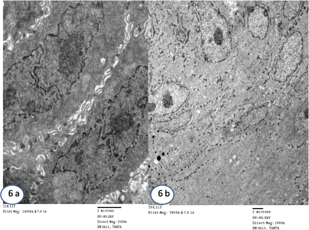
| Figure 6: Electron micrograph of OVX+P group showing: (a) upper layers of prickle cells with multiple mitosis. (Mic. Mag. X 2500). (b) Granular cells showing decreased aggregation of tonofillaments as well as keratohyaline granules. (Mic. Mag. X 1000). |