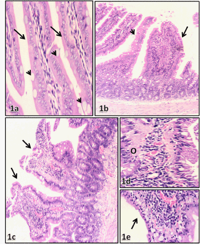
B-E) Transverse sections of duodenum from soriatane treated rats: (b) showed decrease in number of villi, fusion of villi ( arrow), atrophy (head arrow),.H&E,X400. (c) villi stunting (arrows).H&E.X400. (d) edematous (o) of the lamina propria. H&E. X1000. (e) flattening of the epithelial cells with villus core inflammation and villi distortion. H&E. X 1000.