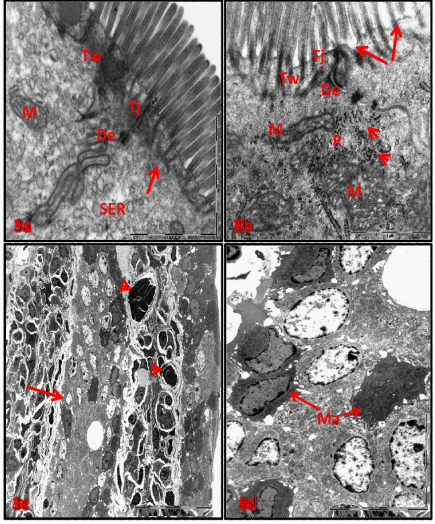
B-D) Electron micrographs of doudenum from soriatane treated rats (b) fusion villi and lose their brush border, enterocytes disorganization (arrow). X850. (c) higher magnification of figure (a) showed macrophages migration (Ma) through mucosa layer. X2600. (d): electron dens necrotic mitochondria(M),fragmentation and degranulation of rough ER (head arrows), increased ribosomal numbers (R), deformed desmosomes (De),terminal web (Tw), tight junction (Tj),with affected microvilli (arrows). X19000.