|
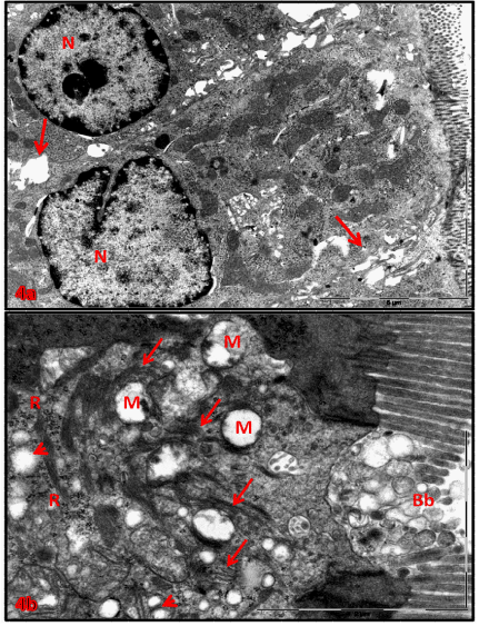
| Figure 4: A, B) Electron micrographs of soriatane treated enterocytes: (a) disruption of lateral infoldings (arrows), and nuclei (N) polymorphism of necrotic enterocytes with increase peripheral chromatin, lose nuclear pores, X5800. (b) microvilli deterioration (Bb), mitochondrial cristiolysis (M), SER proliferation (head arrows), degranulation and atrophied of RER(arrows), increased ribosomal numbers (R). X19000. |