|
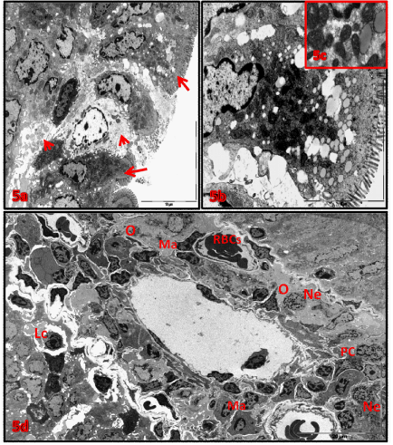
| Figure 5: A-D) Electron micrographs of villi from soriatane treated mucosa: (a) cytoplasmic degeneration (head arrows) and necrotic enterocytes (arrows). X4600. (b) necrotic enterocyte containing electron dens atrophied mitochondria, necrotic nucleus with irregular nuclear membrane and increased number of liposomes. X7900. (c) higher magnification of figure (b) showed atrophied mitochondria and membranous lipid vacuoles. X25000. (d) villus core: showed capillary expanded and congestion, edema(O), increased number neutrophils(Ne), macrophages(Ma), plasma cells(Pc) and lymphocytes (Lc). X1450. |