 |
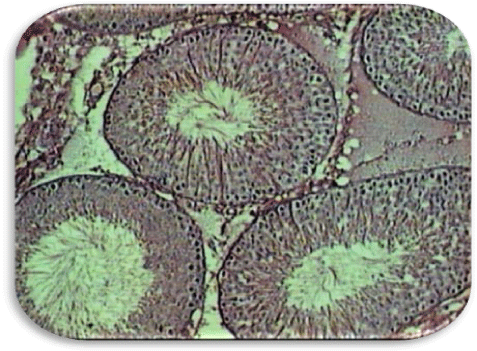
| Figure 1a: Photomicrograph of a cross section of testis from sham operated animal (hematoxylin and eosin staining). Seminiferous tubules showing normal spermatogenesis, exhibiting all stages of spermatogenic cells including abundant spermatozoa. Magnification- 10 X. |