 |
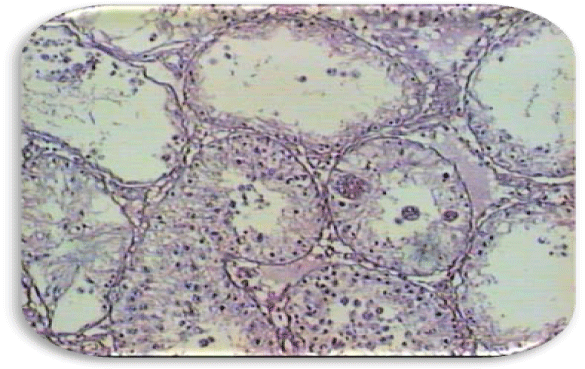
| Figure 1f: Photomicrograph of a cross section from 1 week recovery (hematoxylin and eosin staining). Seminiferous tubules showing sloughing and disorganization. Damage is at the level of spermatozoa, spermatids, spermatocytes and spermatogonia. The interstitial is disorganized and contains some blood. Sertoli’s cells formed multinuclear giant cells. Some tubules are affected at the level of Sertoli’s cells. Magnification 10 X. |