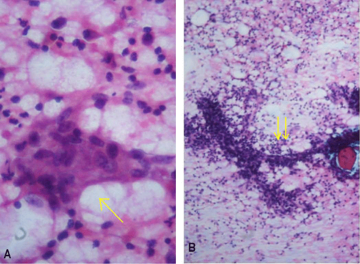
Figure 2:
A. Cytological smear showing an aggregate of epithelioid histiocytes (arrow) within a mixed inflammatory background (Pap x 400). B. Smear showing arborising network of capillary fragments (double arrow) (Pap x400).