|
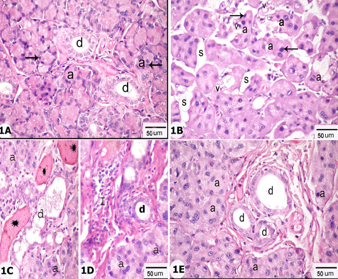
| Figure 1: H&E stained sections of control subgroup Ia (Figure 1A); group II (Figures. 1B,C,D) and group III (Figure 1E): showing the normal serous acini (a) and striated ducts (d) of parotid gland of control group in 1A. Most of the acini of group II have irregular outlines, widely separated (s) and contain darkly stained nuclei (arrows) and vacuoles in Figure (1B). Homogenous acidophilic areas (*) and dilated interlobular ducts (d) are seen in (Figure 1C). Cellular infiltration (I) inside connective tissue septa is prominent in Figure 1D. Figure (1E): showing apparent normal acini and ducts of group III X 400. |