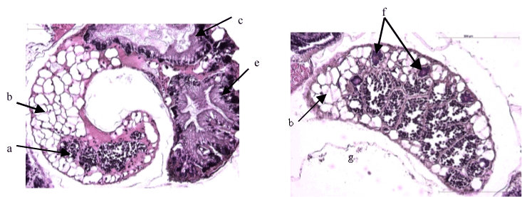
 PLATES 2 and 3 (Figures 8–12): Different gonad maturity stages and internal organization of different organs observed in Valvata piscinalis a) acini; b) albumen
gland; c) digestive tube; d) light of digestive tube; e) digestive gland; f) primary oocytes; g) acini covered with spermatogonia; h) 1st order secondary oocytes;
i) secondary mature oocytes; j) germinating center; k) efferent canal covered with spermatozoids; l) germinating spermatogonia; m) spermatozoids; n) different
maturity stages of spermatogonia, with light increasing along with maturity; o) cells resembling Leydig cells; oc: Leydig cells with Reinke crystalloids; p) intestine;
q) ventricle; q1) atrium; r) embryonic sac; s) nephridial gland; t) mantle; u) columella muscle; v) follicular cord; w) oviduct; x) albumen gland; y) nerve cord; z) mucus
gland; arrow: cells resembling Sertoli cells; Lu: light from the seminal receptacle; *: kidney.
 |
 PLATES 2 and 3 (Figures 8–12): Different gonad maturity stages and internal organization of different organs observed in Valvata piscinalis a) acini; b) albumen
gland; c) digestive tube; d) light of digestive tube; e) digestive gland; f) primary oocytes; g) acini covered with spermatogonia; h) 1st order secondary oocytes;
i) secondary mature oocytes; j) germinating center; k) efferent canal covered with spermatozoids; l) germinating spermatogonia; m) spermatozoids; n) different
maturity stages of spermatogonia, with light increasing along with maturity; o) cells resembling Leydig cells; oc: Leydig cells with Reinke crystalloids; p) intestine;
q) ventricle; q1) atrium; r) embryonic sac; s) nephridial gland; t) mantle; u) columella muscle; v) follicular cord; w) oviduct; x) albumen gland; y) nerve cord; z) mucus
gland; arrow: cells resembling Sertoli cells; Lu: light from the seminal receptacle; *: kidney.