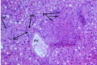
Figure 1:
A photomicrograph of liver section from group 2 showing thickness of portal vein (PV), hepatic artery (HR) and bile ductule (BD), steatosis and foamy vacuoles (FV) with pyknotic stage (PK). (HE, 200X).