|
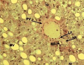
| Figure 4: A photomicrograph of liver section from group 3 showing portal vein (PV) filled with vacuoles, portal triad surrounded by connective tissue and containing hepatic artery (HR) and bile ductule (BD) with infiltrated lipofusin (LF), macro-vacuoles (MV) containing necrosis (Nr) (HE, 200X). |