 |
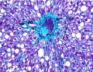
| Figure 9: A photomicrograph of liver section from group 2 showing wall thickness of portal vein (PV) filled with condensed small particles. Collagen fibers (CF) surrounded the portal vein and extended in between the hepatocytes throughout the sinusoids. (Masson trichrome stain, 400X). |