 |
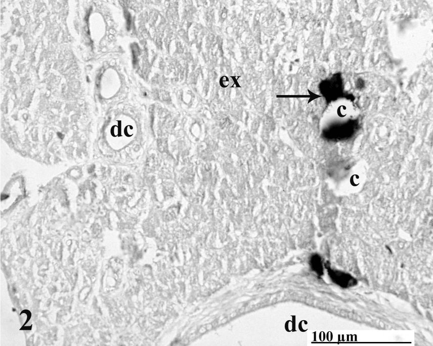
| Figure 2: Succeeding section of preparatory period immunolocalized for glucagon-IR cells (arrow) pouring its secretion into blood capillary (c ). A faint pool of granules moving towards the capillary, smaller and larger ducts (dc) are also seen. X200. |