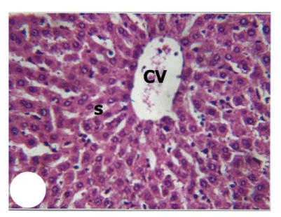
Figure 1:
Light micrograph of a section from normal rat liver (Group1) showing radial organization of hepatocytes and sinusoids (S) around the central vein (CV). [H&E] 100x.