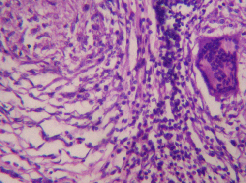
Figure 4:
Photomicrograph of phalanx showing granuloma comprising of circumscribed area with caseating necrosis, epithelioid cells, multinucleated giant cell and peripheral collar of lymphocytes (HE, X400).