|
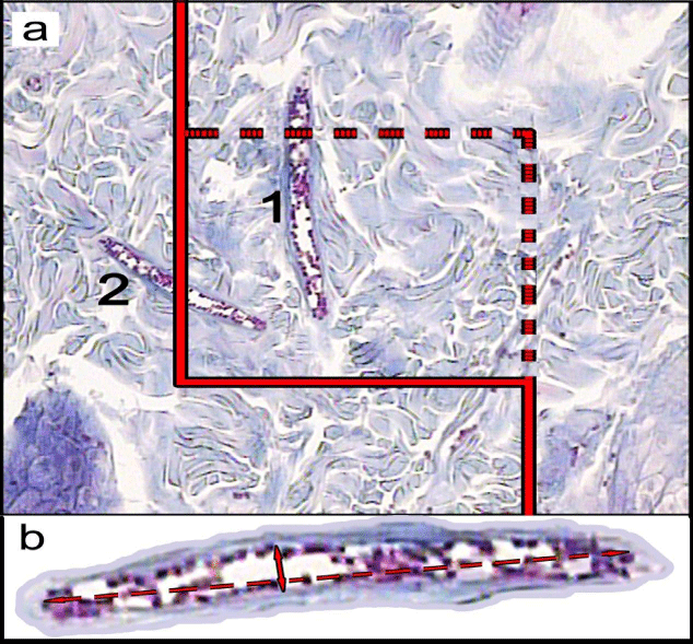
| Figure 4: (a) An unbiased counting frame was laid randomly on the monitor image of wound dermis at final magnification of 450 for estimation of the length density (LV) and mean diameter of the vessels. Any vessel lied in the counting frame (1), or touched the inclusion borders (dotted lines) were selected. The vessels touched the exclusion borders (bold continuous lines), were ignored (2). (b) The short axis of each vessel (short double arrow) was measured as the mean diameter. (Hedenhain’s azan stain; × 450). |