 |
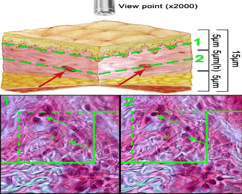
| Figure 5: An unbiased counting frame is superimposed on the monitor image of the 15μm sections to estimate the numerical density (Nv) of the fibroblasts. The traveling height was monitored by a microcator. The range of height that the fibroblasts came into maximal focus was considered as height of disector. (1): The nuclei are unclear at the first 5 μm optical section. (2): As above, any nucleus lied in the counting frame or touched the inclusion borders (green dotted lines) and did not touch the exclusion borders (bold green lines) and come into maximal focus within the next traveling 5 μm optical section (h) are counted (the two arrows). (Hedenhain’s azan stain; × 2000). |