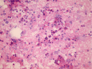
Figure 3:
Smears from subcutaneous nodule in a patient with previous lung SCC show isolated highly atypical keratinized cells with hyperchromatic nuclei in necrotic dirty background. (Pap stain X100).