 |
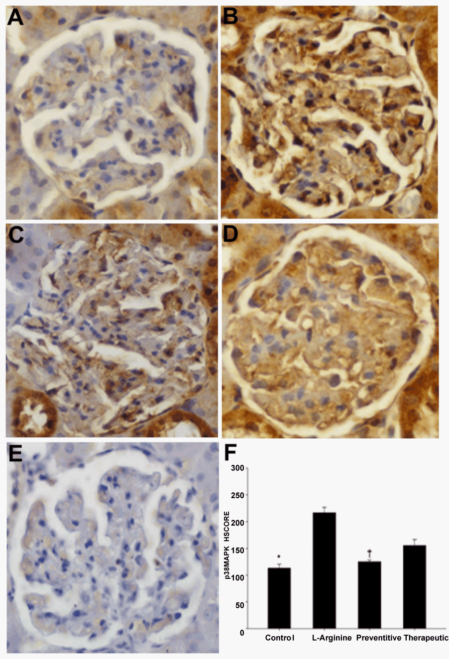
| Figure 1: Immunohistochemical analysis of phospho-p38MAPK expression in control, L-Arginine-given, preventative and therapeutic group mesangial cells. Representative micrographs of serial sections from control (A); L-arginine-given (B); preventative (C) and therapeutic (D) specimens are shown. Phospho-p38MAPK immunostaining (brown) in mesangial cells was mostly cytoplasmic. Between groups, phospho-p38MAPK immunostaining was greater in L-arginine-given (B) vs. control (A) and preventative (C) mesangial cells, while there was no significant difference among L-arginine-given and therapeutic (D) group. Phospho-p38MAPK intensity HSCOREs in control, preventative and L-arginine-given specimens (mean ± SEM) are shown (F); * vs. L-arginine-given and therapeutic mesangial cells; † vs. L-arginine-given and therapeutic mesangial cells; p<0.05. Parallel staining with a rabbit isotype was used as a negative control for phospho-p38MAPK antibody (E). |