|
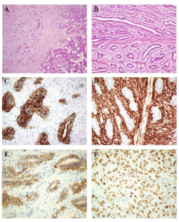
| Figure 3: Histology. a) Normal parotid gland separated from tumor by a connective tissue capsule; H&E, x100. b) Biphasic pattern, an inner ductal cells and outer clear myoepithelial cells; H&E, x200. c) Cytokeratin 8 positivity in the epithelial component; Immunoperoxidase stain, x400. d) Smooth muscle actin positivity in the myoepithelial component; Immunoperoxidase stain, x400. e) CD117 present in the epithelial groups; Immunoperoxidase stain, x400. f) P-63 positivity in the myoepithelial cells nuclei; Immunoperoxidase stain, x600. |