|
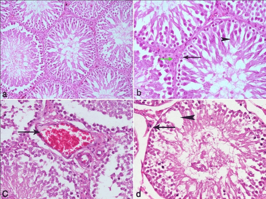
| Figure 1: (a) is a photomicrograph of a section of testis from a control rat showing the normal structure of seminiferous tubules. (b) is a higher magnification of the same section showing the different layers of spermatogenic cells with srtoli cells (arrow) and the old (elongated) spermatids (arrow head) attached to it. In the interstitial tissue a number of Leidig cells (green arrow) are observed. (C) is a photomicrograph of a section of testis from a control radiated rat showing marked dilatation and congestion of blood vessels in interstitial tissue (arrow) with disturbance of spermatogenic layers in many of the tubules and exfoliation of cells in others (arrow head). (d) is a photomicrograph of another section of the same group showing wide gaps in between the spermatogenic cells (arrow head). Atrophy of Leidig cells (arrow) in the interstitial tissue is also observed. (Hx. & E. X 100 & 200). |