|
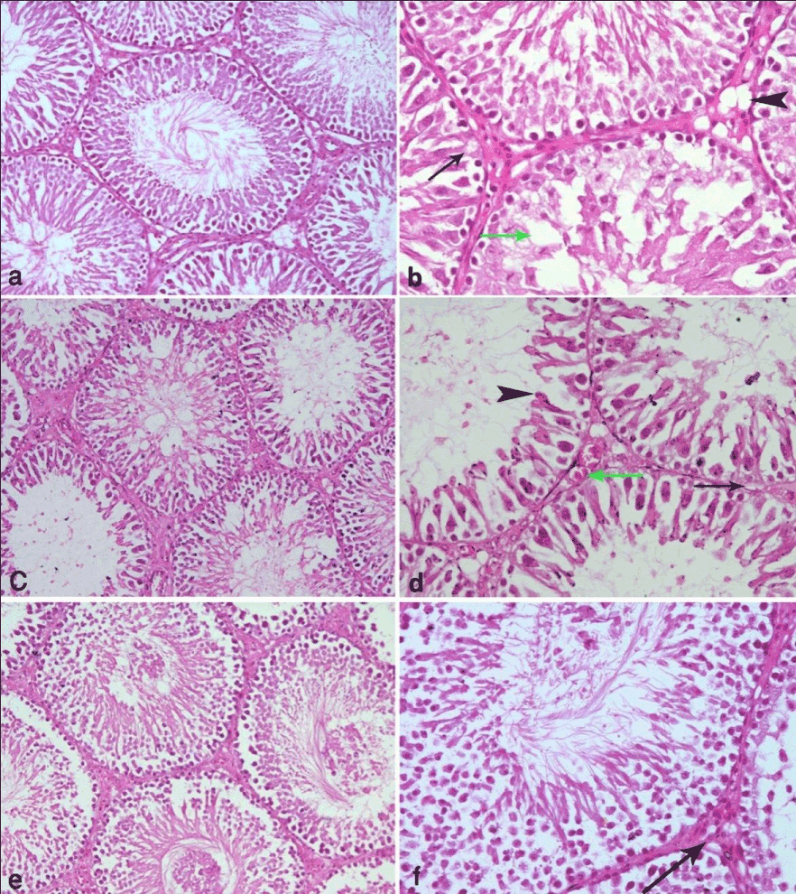
| Figure 2: (a) is a photomicrograph of a section of testis from a rat that received curcumin orally in a dose of 5mg/kg b.w./day for 4 weeks and then radiated with UV radiation in the same dose of previous group showing restoration of the normal structure in most of the seminiferous tubules except for slight vacuolation in the interstitial tissue . (b) is a higher magnification of the same section showing some small vacuoles are still present in the interstitial tissue (arrow head) as well as some small gaps inbetween the spermatogenic cells (green arrow). Sertoli cell with old spermatids attached to it is also observed (arrow). (C) is a photomicrograph of a section of testis from a rat that received curcumin orally in a dose of 25mg/kg b.w./day for 4 weeks and then radiated with UV radiation in the same dose of previous group showing multiple gaps in between the spermatogenic cells (arrow heads) and depletion of spermatogenic cells above the level of spermatocytes in many of the tubules. (d) is a higher magnification of the same section showing no signs of division in spermatocytes, the spermatids are atrophied (arrow head) and the sertoli cells have abnormal-shaped nuclei (arrow). Slight congestion of blood vessels is observed in interstitial tissue (green arrow). (e) is a photomicrograph of a section of testis from a rat that received curcumin orally in a dose of 50 mg/kg b.w./day for 4 weeks and then radiated with UV radiation in the same dose of previous group showing thickening of the basement membrane of seminiferous tubules (arrow heads), exfoliation of spermatogenic cells (arrow) in some tubules and disturbance of spermatobegic layers in others. (f) is a higher magnification of the same section showing depletion of long spermatids and the presence of gaps between the spermatogenic cells (arrow). (Hx. & E. X 100 & 200). |