 |
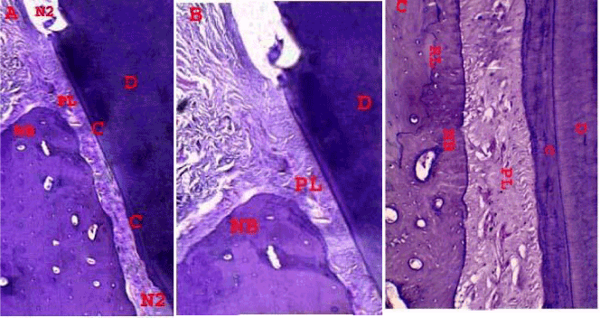
| Figure 3: (a)Histopathology of tested group (strontium ranelate gel) section showing marked newly bone formation (NB) above the notch (N2), Thin layer of acellular cementum at the coronal area of the defect and thick layer at the N2 (H&E . OMX40). (b) High power of tested group shows periodontal ligament with densely arranged good perpendicularly inserted fibers (PL) into the cementum C and newly formed bone( NB) (H&E, OMX100). (c) reversal line was also observed (RL), periodontal ligament with densely well arranged good perpendicularly inserted fibers(PL) into the thick cementum layer near from the N2 (H&E, OMX200). |