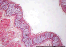
Figure 6:
Photomicrograph of the uterine tube of
P. geoffroanus
showing the nuclei of epithelial cells at different heights (arrow). HE. 100x.