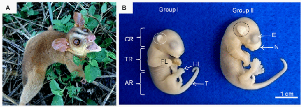
In B: Macroscopic comparison of the developmental stages and external morphological features of foetuses from Groups I and II. Note the division of the main body regions into the cranial region (CR), thoracic region (TR) and abdominal region. In the cranial region, an eye (E) with a pigmented retina, an external ear (circle) and the nasal region (N) can be observed. The forelimbs (FL), hindlimbs (HL) and an elongated tail (T) can also be seen.