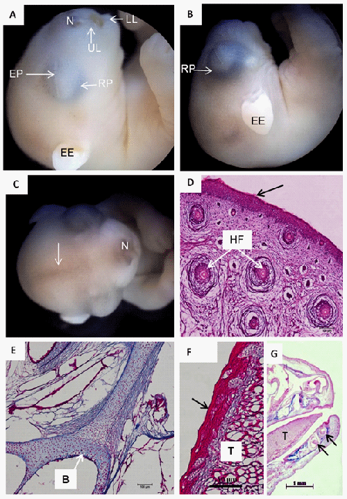
In A: Individual from Group I; and in B-D: Group II. In A: In the oral cavity region, note the upper lip (UL) and lower lip (LL), the short nasal region (N), the eyes with retinal pigmentation (RP), eyelid projection (EP) and a short external ear (EE).
In B-D: Note an increase of the ocular globe and of retinal pigmentation (RP) as well as the length of the external ear (EE). The formation of a darkened strip of hair (arrow) extending from the nasal region (N) to the frontal region.
D: Note an increase in the number of hair follicles (HF) near to the nasal region, as was a thin layer of keratin (arrow) on the dermal epithelium. Staining: hematoxylin and eosin.
In E, Note the ossification process of the bones of the skull base (B) in Group II;
In F, note the keratinised stratified epithelium (arrow) of the tongue (T); and
In G, note the alveolar region of the teeth (arrows). Staining: Masson’s trichrome.