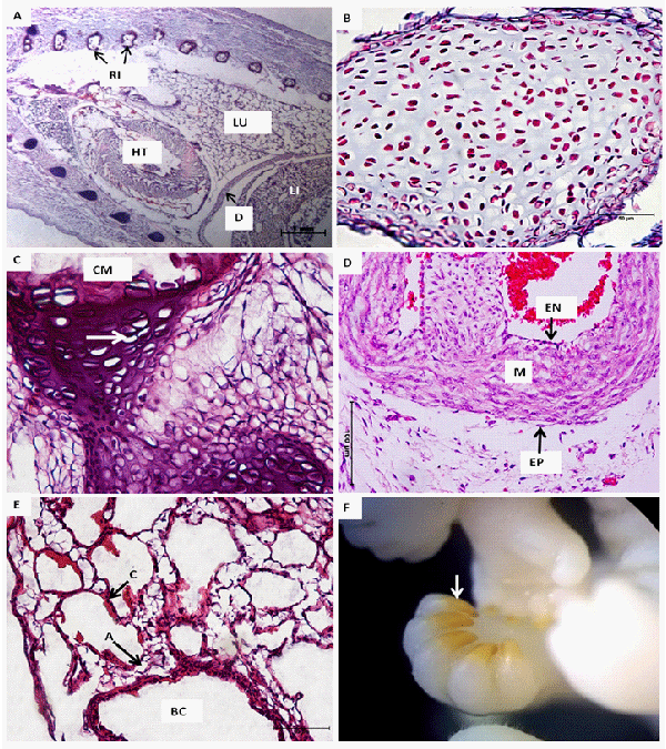
A: Note the syntopy of the heart (HT) and lungs (LU) in the thoracic cavity in an individual from Group I. The ribs (RI) undergoing ossification can also be observed in the cross-sections. The diaphragm (D) separates the thoracic and abdominal cavities. LI = liver.
B: In Group I, the bone moulds are still predominantly formed by cartilaginous tissue.
C: Histological section of the ribs in individual from Group II showing the process of calcification occurring in these bones. Note the mature chondrocytes (arrow) and the region of calcified matrix (CM).
D: In both groups, the heart displayed the three typical layers: the endocardium (EN), myocardium (M) and epicardium (EP).
E: The lungs of both groups consisted of parenchyma with a large number of alveoli (A) in close contact with blood capillaries (C). BC = bronchiole.
F: Detail of the keratinised claws (arrow). Staining: hematoxylin and eosin.