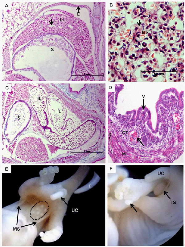
In A: Syntopy of the diaphragm (D) bordering the abdominal cavity, liver (LI) and stomach (S).
B: Detail of the cords of hepatocytes (arrow) surrounded by blood vessels (BV) in the liver parenchyma.
In C: Note the syntopy of the stomach (S) and the intestinal loops (IL), which project into the intestinal lumen, forming the villi.
In D: Note the layers of the intestinal wall formed by the villi (V) of the intestinal epithelium, connective tissue (CT) containing blood vessels (arrow) and muscle layers (M). Staining: hematoxylin and eosin.
In E and F: Differentiation of the external genitalia. In females of both groups, the development of the marsupium (MS) and mammary glands (circle) was noted, and in males, the testicles (TS) were evident in the inguinal region. T = depigmented tail, UC = umbilical cord, and arrow = opposable thumb.