|
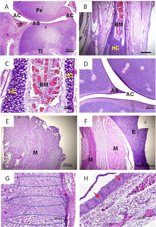
| Figure 2: Development of bone and cartilaginous structures of the pelvic limb in Gallus gallus fetuses at 12 days of incubation and the development of bone and cartilaginous structures of the cranial and cervical region in Gallus gallus fetuses at 14 days of incubation. Bone and articular development. A. Medial view of the stifle joint of 12 days embryos: Femur (FE), Tibia (TI), Articular Surface (AS), Articular Cavity (AC), Articular Capsule (AC). B and C. Long bone (femur): Bone Marrow (BN). D. Articular Surface (AS), Articular Cavity (AC). Note in A, B and C the presence of ossification zone still with a cartilage mold, shown by stars. Observe the skull region of an embryo with 14 days showing the process of endochondral ossification. 20x. G:Photomicrograph of the cervical region of a 14 days embryo, showing the secondary ossification. H: Note that the skull at this stage generally consists of a thin layer of epithelial tissue, corresponding to the skin layers, followed by forming skull and underlying connective tissue, where it is observed a large number of blood vassels. |