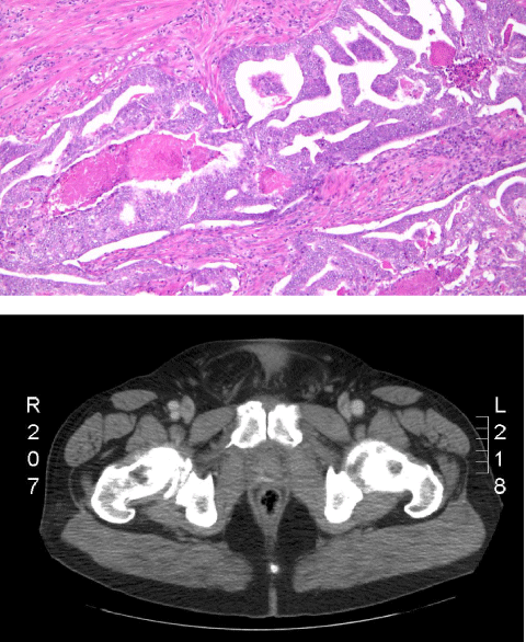
CT Scan pelvis: no evidence of extracapsular extension of tumour
and surrounding basal cells within cribriform structures. Stromal invasion of
surrounding cells corresponding to Gleason’s 4 and areas of comedo necrosis
corresponding to Gleason’s 5.
 CT Scan pelvis: no evidence of extracapsular extension of tumour |
| Figure 2: Ductal adenocarcinoma with pseudostratified columnar epithelium and surrounding basal cells within cribriform structures. Stromal invasion of surrounding cells corresponding to Gleason’s 4 and areas of comedo necrosis corresponding to Gleason’s 5. |