|
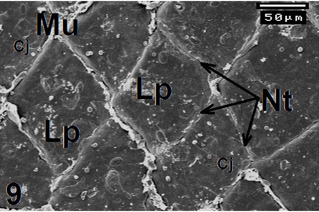
| Figure 9: Electron micrograph shows lingual papillae (Lp) in the middle region of C. ocellatus tongue. Note their rhombus or quadric shapes, the Mucoid secretion (Mu) that emitted from the narrow trenches (Nt) between them. Note the cell junctions between the epithelial cells. ×350. |