|
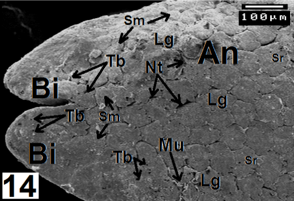
| Figure 14: Electron micrograph shows bi-forked, non-papillosed, keratinized tip (Bi) and large (Lg), multiangular papillae of anterior region of C. sepsoides tongue. Note the serration in the posterior ends of these papillae, the narrow trenches and mucoid secretion that emitted between them. ×150. |