|
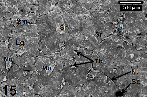
| Figure 15: Electron micrograph shows the dorsal surface of lingual papillae in the middle region of C. sepsoides tongue. Most of the lingual papillae are large (Lg) with few smaller ones (Sm). Note the serrated ends (Sr), secretory orifice (So) and taste bud (Tb) orifices. ×350. |