|
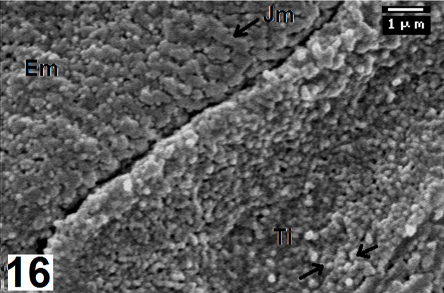
| Figure 16: Magnified micrograph from figure 15 shows the microvilli that cover epithelial cells in the serrated area of a lingual papilla. Upper left part provided with thick, enlarged microvilli (Em), which may fused together to form jagged microvilli (Jm) that are connected with each other by microridges (Mr). Lower right shows tiny microvilli (Ti), some of them have spherical terminus (arrows). ×10,000. |