|
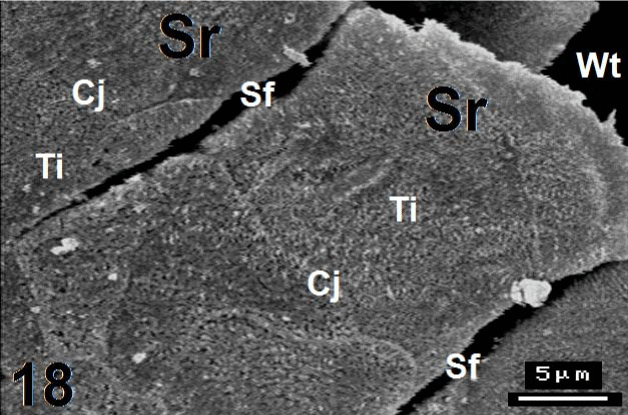
| Figure 18: Magnified micrograph from figure 17 shows the serrated (Sr) part of a lingual papilla in the posterior region of the tongue. Two serration furrows (Sf) are dividing the terminal end of the present lingual papilla. Note the cell junctions (Cj) between them and their tiny microvilli (Ti). Wt: wide trench. ×3500. |