 |
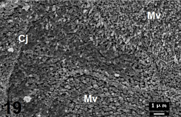
| Figure 19: Magnified micrographs from figure 17 shows different shapes of microvilli (Mv) in the posterior region of C. sepsoides tongue. Figure 19 shows loosely arranged (upper right) or aggregated (lower middle region) microvilli. Note the cell junctions (Cj) between the epithelial cells. ×7500. |