 |
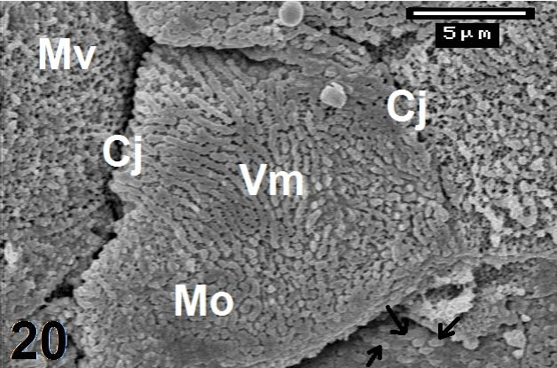
| Figure 20: Magnified micrographs from figure 17 shows different shapes of microvilli (Mv) in the posterior region of C. sepsoides tongue. Figure 20 displays other diffferent patterns of microvilli in the cerrated portion of lingual papilla. The central epithelial cell is studded by a condensed architectural pattern of mosaic (Mo) and virtuous microvilli (Vm) with narrow spaces are separating between them. Left epithelial cell shows that the microvilli (Mv) have fine cylindrical shapes with spherical terminus, while that in the right are provided microvilli with large spherical terminus (arrow). Cj: Cell junction. ×5000. |