 |
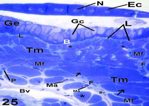
| Figure 25: Light micrograph shows transverse semithin section in the anterior region of C. ocellatus tongue. The upper side shows a single layer of epithelial cells (Ec) with their central nuclei (N) at the posterior extremity of a lingual papilla. The lower side shows underneath papilla formed of glandular epithelium (Ge), thick basement membrane (B), lamina propria (L), tongue muscles (Tm). The muscle fibers (Mf) are cut in a transverse (stars) and longitudinal plan (arrows). Note the presence of some blood vessels (Bv), numerous fibrocytes (F), few mast cells (Ma) and plasma cells (P). ×160. |