|
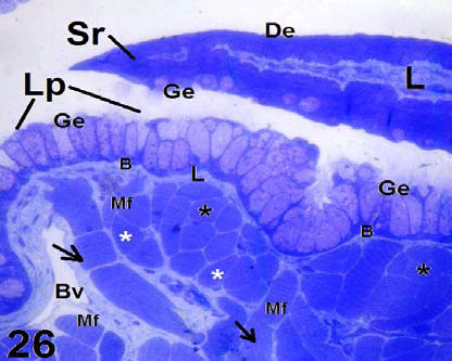
| Figure 26: Light micrograph shows longitudinal semithin section in the posterior region of C. ocellatus tongue. The upper side shows posterior serrated (Sr) part of a ligual papilla. It is formed of dorsal epithelium (De), lamina propria (L) and ventral glandular epithelium (Ge) . The lower side shows the an anterior part of another lingual papilla follower the previous one. Note the basement membrane (B), Blood vessels (Bv). The muscle fibers (Mf) are cut in a transverse (stars) and vertical (arrows) plan. ×160. |