 |
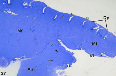
| Figure 27: Light micrograph shows longitudinal semithin section of C. sepsoides tongue. The dorsal papillae (Dp) is a large, flattened and are separated by narrow trenches (Nt). Most of them are imbricated (Im) at their anterior ends. The ventral lingual epithelium (Vl) contains numerous lingual folds (Lf). The lingual connective tissue rich with lingual muscle fibers (Mf) more than that, described in C. ocellatus tongue. Note that the tongue is supported by an accessory lingual muscle (Am), which may increase potency of tongue movements. This muscle is supported by muscle fibers (Mf) that was cut in longitudinal (Lon), Transverse (Tr) and vertical plans (V). ×25. |