 |
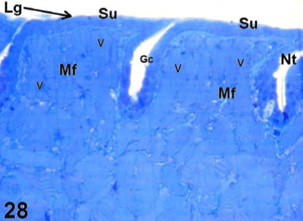
| Figure 28: Light micrograph magnified from 27 shows large lingual papillae (Lg) of the anterior lingual region, surface epithelium (Su), narrow trenches (Nt) and few goblet cells (Gc). Note that each papilla is supplied by large number of muscle fibers (Mf) that are cut in a vertical plan to facilitate expansion of lingual papillae and protrusion of the tongue. ×100. |