 |
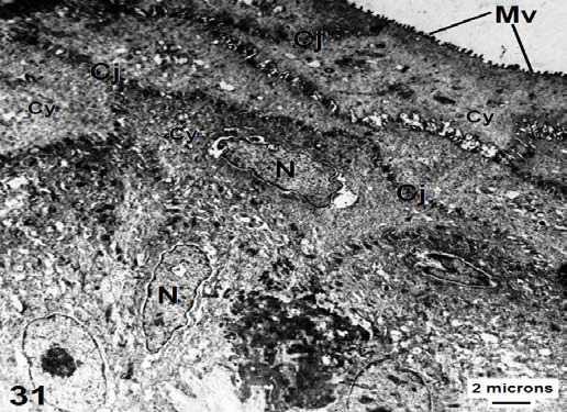
| Figure 31: Transmission electron micrograph shows stratified squamous keratinized epithelium of the tip region of C. sepsoides tongue. Note the cell junctions (CJ), microvilli (Mv), their electron dense cytoplasm (Cy) and elongated nuclei (N). ×6000. |