 |
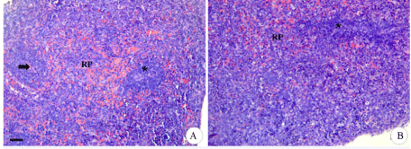
| Figure 4: Photomicrographs of spleen of neonatal mice euthanized after 24 hours of life. A) CG with visible delineation of spleen pulps. It is noticed formation of white saves around the central arteriole (arrow). B) ICS21 showing undefined boundary between white pulp (asterisk) and red pulp (RP). H and E. Bar: 20 μm. |