 |
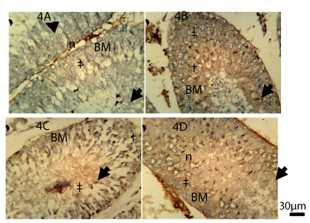
| Figure 4: CD3+ immunohistochemistry (for undifferentiated lymphocytes) in the germinal epithelium of wild type adult Sprague-Dawley rats. (A) Control (B) 100mg/Kg Pb-Acetate (C) Pb-Acetate+Se+Zn and (D) Se+Zn only. CD3+ activity is high in the BM of the Pb treatment (4B), in the BM and degenerating cells of the lead treated group (4B), in the BM of Pb+Se+Zn and it is widely diffused in the epithelial cells of the Se+Zn treated group (4C and 5D). (‡) represents regions of CD3+ immunopositivity and (†) represents regions of cellular degeneration, (n) represents normal cells of the epithelium, (BM) basement membrane, arrow head indicates the lumen of the seminiferous tubule (Scale bar is 30 μm). |