|
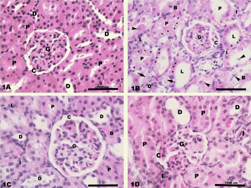
| Figure 1: Light photomicrographs of rats renal cortex showing 1A) normal structure in control rats; 1B) degeneration and necrosis (*) of the proximal tubular cells (P) in cisplatin-treated rats. Both proximal and distal (D) tubules contain detached epithelial cells (arrow head) and excessive amount of cast (arrow) within their wide lumina (L). The renal corpuscle shows condensation of the glomerular capillary tuft (G) and dilation of the capsular space (C). Peritubular cellular infiltration (I) and congestion (B) are seen as well; 1C) The renal cortex shows an interstitial cellular infiltration (I) and congestion (B) in the misoprostoltreated rats; 1D) A few degenerated epithelial cells (*) are seen in few proximal tubules (P) with peritubular cellular infiltration (I) and congestion (B) in combined misoprostol and cisplatin- treated rats. |