|
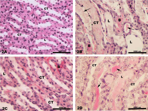
| Figure 2: Light photomicrographs of rats renal medulla showing 2 A) the collecting tubules (CT) having normal structure and empty lumen (L) in control rats; 2B) the collecting tubules (CT) have thin necrotic wall (*) and dilated lumen (L) containing an excessive amount of cast deposition (arrow) in cisplatin-treated rats. Also, peritubular cellular infiltration (I) and congestion (B) are seen within the renal medulla in cisplatin-treated rats as well; 2C) the collecting tubules (CT) show normal structure with peritubular congestion (B) in misoprostol-treated rats; 2D) homogenous cast (arrow) is seen within the slightly dilated lumen of the collecting tubules (CT) in combined misoprostol+ cisplatin-treated rats. |