|
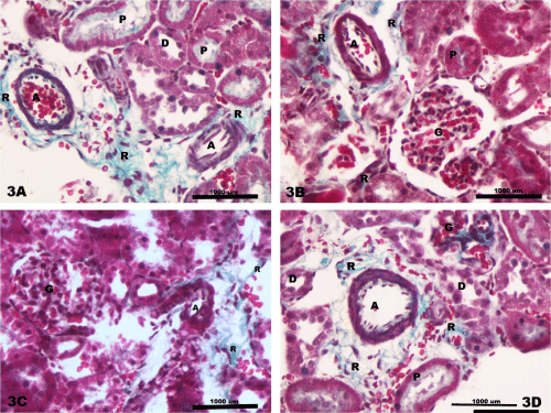
| Figure 3: Light photomicrographs of masson trichrome stained sections of rat renal cortex showing little amount of the collagen fibers (R) distributed mainly around the renal vascular structures (A) in control rats (3A), cisplatin-treated (3B), misoprostol-treated (3C) and combined misoprostol and cisplatin-treated (3D) rats with no noticeable difference. |