 |
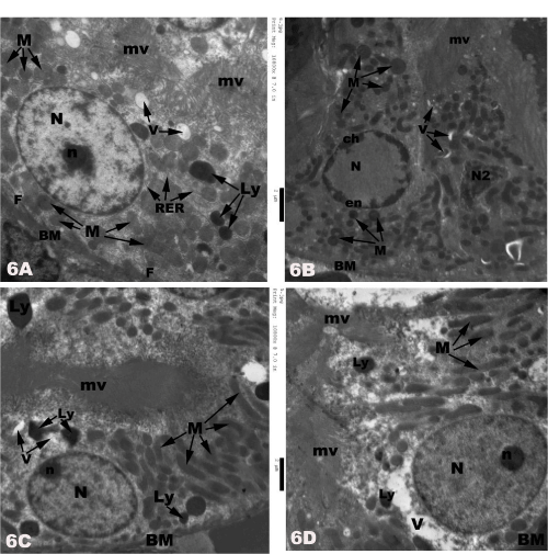
| Figure 6: Electron micrographs of rats proximal tubular cells showing 6A) in control rats, basal infolding (F) extending from the regular basement membrane (BM) into the cellular cytoplasm. The cell has large oval basal nucleus (N) with central nucleolus (n). Its cytoplasm contains many round-shaped normal basal mitochondriae (M), different sized scattered lysosomes (Ly), few short rough endoplasmic reticulum cisternae (RER) and apical electron-lucent vacuoles (V). Numerous long microvilli (mv) are seen at the luminal side of the cell; 6B) in cisplatin-treated rats, marked reduction of the cells size and different apoptotic nuclear changes (N2) with margination of the heterochromatin (ch) on the inner aspect of the nuclear envelope (en) are seen in many cells. Also, thickness of cell basement membrane (BM) with absence of its basal infoldings, many smallsized condensed mitochondriae (M) with different shapes and fragmentation of the apical microvilli (mv) are noticed as well. 5C) the proximal tubular cell shows basal oval euchromatic nucleus (N) with peripheral nucleolus (n), many round and elongated basal mitochondria (M), few lysosomes (Ly) and numerous long microvilli (mv) in misoprostol-treated rats; 6D) normal basement membrane (Bm), large-sized oval basal euchromatic nucleus (N)with peripheral nucleolus (n), electron-lucent vacuoles (V), few apical lysosomes (Ly)and long numerous microvilli are observed in the proximal tubular cell in combined misoprostol and cisplatin-treated rats. |