|
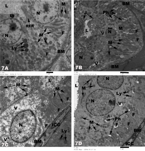
| Figure 7: Electron micrographs of rats distal tubular cells showing 7A) normal cells with regular basement membrane (BM) and many basal infolding (F). The cells contain numerous normal elongated basal mitochondriae (M), many basal lysosomes(Ly), few short rough endoplasmic reticulum cisternae (RER) and apical round euchromatic nucleus N) with central nucleolus (n). Well delineated apical cell membrane without microvilli and wide tubular lumen (L) are seen in the control rats; 7B) thick basement membrane (BM) with increase number of basal infolding (F), nuclear degeneration (N) and fragmentation of nuclear envelope (ne), presence of many phagolysosomes (Ph), scattered vacuoles (V) and degeneration and reduction of mitochondrial number (M) and apical tight junctions (J) between the cells are seen in cisplatin-treated rats; 7C) normal basement membrane (BM) with many basal infoldings (F), many elongated basal mitochondriae (M), central oval euchromatic nucleus (N), few electronlucent vacuoles (V) and apical tight junctions (J) with presence of intercalated cell (ICC) between the distal tubular cells are observed in misoprostol-treated rats; 7D) normal basement membrane (BM) with many basal infoldings (F) and intercalated cell (ICC) are seen in the combined misoprostol and cisplatin-treated rats. Also, numerous elongated normal basal mitochondriae (M), few lysosomes (Ly) and phagosomes (Ph), basal vacuoles (V) and apical round euchromatic nucleus (N) with peripheral nucleolus (n) are seen as well. |